Types of Electron Microscopes
| Home | | Pharmaceutical Microbiology | | Pharmaceutical Microbiology |Chapter: Pharmaceutical Microbiology : Identification of Microorganisms
The electron microscopes are of two different types, namely : (a) Transmission Electron Microscope (TEM), and (b) Scanning Electron Microscope (SEM).
Electron Microscope
An electron microscope refers to a microscope that makes use of streams of electrons duly deflected from their course either by an electrostatic or by an electromagnetic field for the magnifica-tion of objects. The final image is adequately viewed on a fluorescent screen or recorded on a photo-graphic plate. By virtue of the fact that an electron microscope exhibits greater resolution, the ensuing images may be magnified conveniently even upto the extent of 4,00,000 diameters.
It is, however, pertinent to mention here that objects that are smaller than 0.2 μm, for instance : internal structures of cells, and viruses should be examined, characterized and identified by the aid of an electron microscope.
Importantly, the electron microscope utilizes only a beam of electrons rather than a ray of light. The most acceptable, logical, and plausible explanation that an electron microscope affords much prominent and better resolution is solely on account of the fact that the ‘electrons’ do possess shorter wavelengths significantly. Besides, the wavelengths of electrons are approximately 1,00,000 times smaller in comparison to the wavelengths of visible light.
Interestingly, an electron microscope predominantly employs electromagnetic lenses, rather than the conventional glass lenses in other microscopes ; and ultimately, focused upon a ‘specimen’ a beam of electrons which is made to travel via a tube under vacuum (so as to eliminate any loss of energy due to friction collision etc.).
Types of Electron Microscopes :
The electron microscopes are of two different types, namely :
(a) Transmission Electron Microscope (TEM), and
(b) Scanning Electron Microscope (SEM).
These two types of electron microscopes shall be discussed briefly in the sections that follows :
(a) Transmission Electron Microscope (TEM)
The transmission electron microscope (TEM) specifically makes use of an extremely fine focused beam of electrons released precisely from an ‘electron gun’ that penetrates via a specially prepared ultrathin section of the investigative specimen, as illustrated in Fig. 4.9.
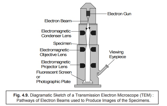
Methodology : The various operational steps and vital components of TEM are described below :
(1) The Electron Gun i.e., a pre-heated Tungsten Filament, usually serves as a beam of elec-trons which is subsequently focussed upon the desired ‘specimen’ by the help of Electromagnetic Condeser Lens.
(2) Because the electrons are unable to penetrate via a glass lens, the usage of doughnut-shaped electromagnets usually termed as Electromagnetic Objective Lenses are made so as to focus the beam properly.
(3) The entire length of the column comprising of the vairous lenses as well as specimen should be maintained under high vacuum*.
(4) The specimen causes the scattering of electrons that are eventually gaining an entry through it.
(5) The ‘electron beam’ thus emerged is adequately focused by the aid of Electromagnetic Projector Lenses strategically positioned which ultimately forms an enlarged and distinctly visible image of the ‘specimen’ upon a Fluorescent Screen (or Photographic Plate).
(6) Specifically the appearance of a relatively denser region in the ‘specimen’ helps to scatter much more electrons ; and, hence, may be viewed as darker zones in the image because only fewer electrons happen to touch that particular zone of the fluorescent screen (or photographic plate).
(7) Finally, in a particular contrast situation, the electron-transparent zones are definitely brighter always. The ‘screen’ may be removed and the image may be captured onto a ‘photographic plate’ to obtain a permanent impression as a record.
(b) Scanning Electron Microscope (SEM)
As it has been discussed under TEM that an image can be obtained from such radiation which has duly transmitted through a specimen. In a most recent technological advancement the Scanning Electron Microscope (SEM) has been developed whereby detailed in-depth examination of the sur-faces of various microorganisms can be accomplished with excellent ease and efficiency. In reality, the SEM markedly differs from several other electron microscopes wherein the image is duly obtained right from the electrons that are strategically emitted by the surface of an object in comparison to the transmitted electrons. Thus, there are quite a few SEMs which distinctly exhibit a resolution of 7 nm or even less.
Fig. 4.10. duly depicts the diagramatic sketch fo a Scanning Electron Microscope (SEM) that vividly shows the primary electrons sweeping across the particular investigative specimen together with the knock electrons emerging from its surface. In actual practice, these secondary electrons (or knock electrons) are meticulously picked up by a strategically positioned collector, duly amplified, and transmitted onto a viewing screen or a photographic plate (to have a permanent record/impression of the investigative specimen).
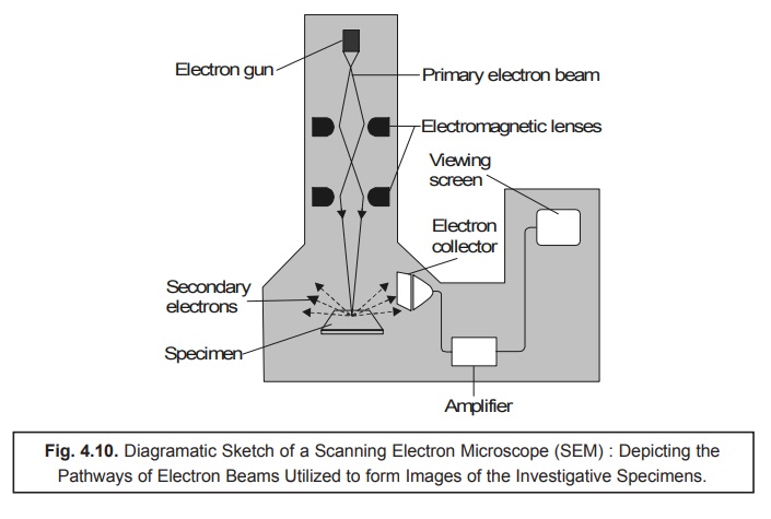
Methodology : The various steps involved in the operative sequential steps are as stated under :
(1) Specimen preparation : It is quite simple and not so cumbersome ; and even in certain cases one may use air-dried specimen for routine examination directly. In general, largely the microor-ganisms should first be fixed, dehydrated, and dried meticulously so as to preserve not only the so called ‘surface-structure’ of the specimen but also to prevent the ‘possible collapse of the cells’ when these are directly exposed to the SEM’s high vacuum. Before, carrying out the usual viewing activities, the dried samples are duly mounted and carefully coated with a very thin layer of metal sheet in order to check and prevent the buildup of an accumulated electrical charge onto the surface of the specimen and to provide a distinct better image.
(2) SEM helps in the scanning of a relatively narrow and tapered electron beam both back and forth onto the surface of the specimen. Thus, when the beam of electron happens to strike a specific zone of the specimen, the surface atoms critically discharge a small shower of electrons usually termed as ‘secondary electrons’, which are subsequently trapped and duly registered by a specially designed detector.
(3) The ‘secondary electrons’ after gaining entry into the ‘detector’ precisely strike a scintillator thereby enabling it to emit light flashes which is adequately converted into a stream of elctrical current by the aid of a photomultiplier tube. Finally, the emerging feeble electrical current is duly amplified.
(4) The resulting signal is carefully sent across to a strategically located cathode-ray tube, and forms a sharp image just like a television picture, that may be either viewed or photographed accord-ingly for record.
Notes :
(i) The actual and exact number of secondary electrons that ultimately reach the ‘detector’ exclu-sively depends upon the specific nature of the surface of the investigative specimen.
(ii) The ensuing ‘electron beam’ when strikes a raised surface area, a sizable large number ‘sec-ondary electrons’ gain due entry into the ‘detector’ ; whereas, fewer electrons do escape a depression in the surface of the specimen and then reach the detector. Therefore, the raised zones appear comparatively lighter on the screen, and the depressions are darker in appear-ance. Thus, one may obtain a realistic 3D image of the surface of the microorganism having a visible intensive depth of focus.
Related Topics