T-Lymphocyte Antigen Recognition and MHC Proteins
| Home | | Pharmaceutical Microbiology | | Pharmaceutical Microbiology |Chapter: Pharmaceutical Microbiology : Immunology
Epitopes for T-lymphocytes comprise exclusively linear peptide sequences. T-lymphocytes are unable to respond to carbohydrate, lipid or nucleic acid material and they only respond to peptide antigen........
T-LYMPHOCYTE ANTIGEN RECOGNITION AND MHC
PROTEINS
Epitopes for T-lymphocytes comprise
exclusively linear peptide sequences. T-lymphocytes are unable to respond to
carbohydrate, lipid or nucleic acid material and they only respond to peptide
antigen when it is presented to the T-lymphocyte by surface proteins on the
plasma membrane of host cells. These surface proteins are termed MHC proteins
and can be subdivided into two main classes:
• MHC class I proteins are expressed on the surface of all nucleated
host cell membranes and present peptide antigen to cytotoxic T-lymphocytes.
• MHC class II proteins are expressed only on a more specialized
group of cells termed antigen-presenting cells (APCs), and present peptide
antigen to helper T-lymphocytes.
Such a distinct cellular distribution of
MHC proteins and restriction in presentation to discrete T-lymphocyte
sub-populations may be remembered by considering the different T-lymphocyte
functions. That is, all cells of the body have the potential to become infected
with virus and undergo a cancerous change, and hence all cells must have the
capacity to be destroyed by the actions of cytotoxic T-cells. As such, all
cells of the body must possess MHC I molecules to afford antigen presentation
to cytotoxic T-cells. In contrast, as a coordinator cell of the immune system, the
helper T-cell must be able to respond to its environment in order to give
appropriate signals or ‘help’ to other immune cells. Specialized APCs with the
capacity to phagocytose interstitial proteinaceous material therefore undertake
the function of ‘environmental sampling’. MHC class II proteins expressed on
the surface of APCs will present peptide antigen to helper T-lymphocytes.
APCs include the macrophage tissue cell
population, specialized APCs such as dendritic cells within the lymphatic
system or Langerhans cells within the skin. B-lymphocytes also serve the
function of an APC because they interact with protein antigen through
high-affinity surface IgM molecules. Subsequently, they internalize the protein
antigen for processing to generate peptides that will be presented by MHC II
molecules expressed on the B-lymphocyte cell surface. Another cell type that
can serve the function of an APC is the endothelial cell, which can be induced
to express MHC II molecules by the action of the cytokine IFN-γ. It should not
be overlooked that the APC can itself become infected with virus and undergo
cancerous transformation, and therefore the APC, in addition to MHC II
molecules, will also express the full complement of MHC I molecules on its
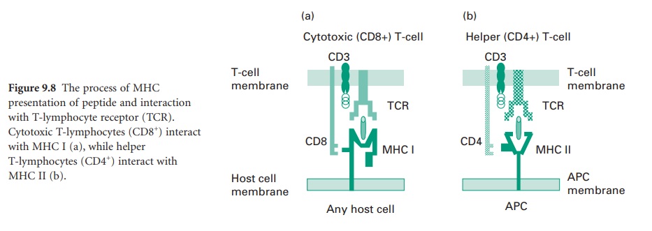
surface. This process of MHC presentation of peptide and interaction with
T-lymphocyte receptor is shown in Figure 9.8. A foreign peptide presented by a
MHC molecule will be recognized by a TCR expressed on the surface of an
appropriate T-lymphocyte. Once a particular TCR recognizes a peptide sequence
as foreign, intracellular signals to activate the T-cell are sent via the CD3
complex present within the T-cell membrane. The recognition of peptide as
foreign will lead to an immune response. Beyond antigen presentation, the interaction
between MHC molecule and T-lymphocyte also serves to identify that the
T-lymphocyte and host cell membrane arise from the same embryonic tissue.
Tremendous interindividual differences, or more specifically polymorphisms,
exist in the MHC proteins within a population. The T-lymphocyte undertakes this
MHC surveillance through the possession of accessory molecules. Cytotoxic
T-lymphocytes possess CD8+ molecules which interact with MHC I (Figure 9.8a),
while helper T-lymphocytes possess CD4+ molecules which interact with MHC II
(Figure 9.8b); hence the use of the terms CD8+ lymphocytes to refer to
cytotoxic T-cells and CD4+ lymphocytes to refer to helper T-cells.
Related Topics
