Organization of Microbial Cells
| Home | | Pharmaceutical Microbiology | | Pharmaceutical Microbiology |Chapter: Pharmaceutical Microbiology : Structure and Function of Bacterial Cells
Irrespective of the very nature and complexity of an organism, the cell designates the fundamen-tal structural unit of life. In other words, all living cells are basically similar.
ORGANIZATION
OF MICROBIAL CELLS
Irrespective
of the very nature and complexity of an organism, the cell designates the
fundamen-tal structural unit of life. In other words, all living cells are
basically similar. The ‘cell theory’
i.e., the concept of the cell as the
structural unit of life, was duly put forward by Schleiden and Schwann.
In short,
all biological systems essentially have the following characteristic features
in common, namely :
(1) ability
for reproduction,
(2) ability
to assimilate or ingest food substances, and subsequently metabolize them for
energy and growth,
(3) ability
to excrete waste products,
(4) ability
to react to unavoidable alterations in their environment — usually known as irritabil-ity, and
(5) susceptibility
to mutation.
The plants and the animals were the two
preliminary kingdoms of living organisms duly rec-ognized and identified by the
earlier biologists. However, one may
articulately distinguish these two groups by means of a number of well-defined
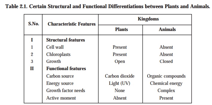
structural and functional characteristic features as given in Table : 2.1.
Soon
after the discovery of the ‘microbial
world’ — the immotile multicellular and photosyn-thetic algae were
classified duly in the plant kingdom;
whereas — the microscopic motile forms of algae were placed duly in the animal kingdom. Hence, a close and
careful examination revealed the presence of both plant — and animal-like
characteristic features in the ‘microorganisms’.
Further, supporting evidences and valuable informations strongly established
that the ‘microorganisms’ could not
fit reasonably into the two aforesaid kingdoms, namely : ‘plants’ and ‘animals’.
Therefore, Haeckel (1866) legitimately and affirmatively proposed a ‘third kingdom’ termed as the ‘Protista’* to include the ‘microorganisms’ exclusively.
Importantly,
Protista group usually comprises of
both the photosynthetic and non-photosyn-thetic microorganisms, of
course, with certain members sharing the characteristic features of both the usual traditional plant and animal
kingdoms. Nevertheless, the most prominent and predominant at-tribute of this
particular group being the comparatively much simpler biological organization.
Most of the representative members of this group are normally unicellular and
undifferentiated unlike the ani-mals and the plants.
Noticeably,
further categorization of this kingdom was exclusively dependent upon the
extent of complexity encountered by the cellular organization, substantial
progress in microscopy, and the ‘bio-chemistry of various microorganisms’ has
ultimately paved the way towards a much advanced and better understanding of
the differences with regard to the ‘internal
architectural design of the micro-bial cells’.
Types of Cells
As to
date there are two types of cells
that have been recognized duly, such as :
(a) Eukaryotic
cells, and
(b) Prokaryotic
cells.
Another
third type, known as the Urkaryotes,
and are most probably the progenitor of the present day eukaryotes has now also been recognized duly.
The above
two types of cells* (a) and (b) shall now be discussed at length in the sections that follows :
(a) Eukaryotic
Cells [‘eu’ = true ; ‘karyote’ = nut (refers to nucleus of cell)] ;
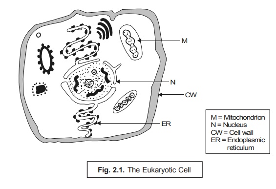
It has
been observed that the eukaryotic cells
(Fig. 2.1) are explicitely characterized by the presence of a multiplicity of definite unit membrane
systems that happen to be both structurally and topologically distinct from
the cytoplasmic membrane. Subsequently, these prevailing membrane sys-tems
categorically enable the segregation of
various eukaryotic cytoplasmic functions directly into specialized organelles.**
Endoplasmic reticulum (ER) represents the most complex internal mem-brane
system that essentially comprises of an irregular network of interconnected
delimited channels that invariably cover a larger segment of the interior
portion of the cell. Besides, ER gets in direct contact with two other extremely vital components viz., nucleus and cytoplasmic
ribosomes. The nu-cleus membrane is duly formed by a portion of the endoplasmic reticulum surrounding the
nucleus ; whereas, in other regions the surface of the membrane is particularly
covered with the ribosomes wher-ever synthesis of protein takes place. The
proteins thus generated eventually pass via
the endoplasmic reticulum channels right to the various segments of the ensuing cell cytoplasm.
Nucleus. The eukaryotic cell possesses the
‘genetic material’ duly stored in the chromosomes i.e., very
much within the nucleus. However,
chloroplasts and mitochondria also
comprise of character-istic DNA. The
chromosomes are linear threads made of DNA (and proteins in eukaryotic cells)
in the nucleus of a cell, which may stain deeply with basic dyes, and are found
to be especially conspicuous during mitosis. The DNA happens to be the genetic
code of the cell ; and specific sequences of DNA nucleotides are the genes for
the cell’s particular proteins. However, the size and the number of the
chromosome vary widely with various organisms. Nevertheless, the nucleus
invariably contains a nu-cleolus that
is intimately associated with a particular chromosomal segment termed as the ‘nucleolar organizer’,
which is considered to be totally involved in ribosomal RNA (rRNA)
synthesis.
Mitosis. Mitosis refers to a type of cell division
of somatic cells wherein each daughter cell
contains the same number of chromosomes as the parent cell. Mitosis is the specific process by
which the body grows and dead somatic cells are replaced. In fact, mitosis is a continuous process divided
into four distinct phases, namely : prophase, metaphase, anaphase, and telophase.
A brief
discussion of the aforesaid four
phases shall be given in the sections that follows along with their
illustrations in Fig. 2.2.
(a) Prophase. In prophase, the chromatin granules of the nucleus usually stain more
densely and get organized into chromosomes. These first appear as long
filaments, each comprising of two
identical chromatids,* obtained as a
result of DNA replication. As prophase
progresses, the chromosomes become shorter and more compact and stain densely.
The nuclear mem-brane and the nucleoli disappear. At the same time, the centriole divides and the two daugh-ter centrioles,** each
surrounded by a centrosphere, move to
opposite poles of the cell. They are
duly connected by fine protoplasmic fibrils, which eventually form an achromatic spindle.
(b) Metaphase. The metaphase refers to the chromosomes
(paired chromatids) that arrange themselves in an equatorial plane midway
between the two centrioles.
(c) Anaphase. In anaphase, the chromatids
(now known as daughter chromosomes) diverge and move towards their respective centrosomes. The end of their migration
marks the begin-ning of the next phase.
(d) Telophase. In telophase, the chromosomes at each pole of the spindle undergo
changes that are the reverse of those in the prophase, each becoming a long
loosely spiraled thread. The nuclear membrane re-forms and nucleoli reappear.
Outlines of chromosomes disappear, and chromatin appears as granules scattered
throughout the nucleus and connected by a highly staining net. The cytoplasm
gets separated into two portions,
ultimately resulting in two complete
cells. This is accomplished in animal cells by constriction in the equatorial
region ; in plant cells, a cell plate that produces the cell membrane forms in
a similar position. The period between two successive divisions is usually
known as interphase.
Mitosis is of particular significance
wherein the genes are distributed equally to each daughter cell and a fixed number of chromosomes is maintained in all somatic
cells of an organism.
Mitosis are of two kinds, namely :
(i) heterotypic mitosis : The
first or reduction division in the maturation of germ cells, and
(ii) homeotypic
mitosis : The second or equational division in the maturation of germ
cells.
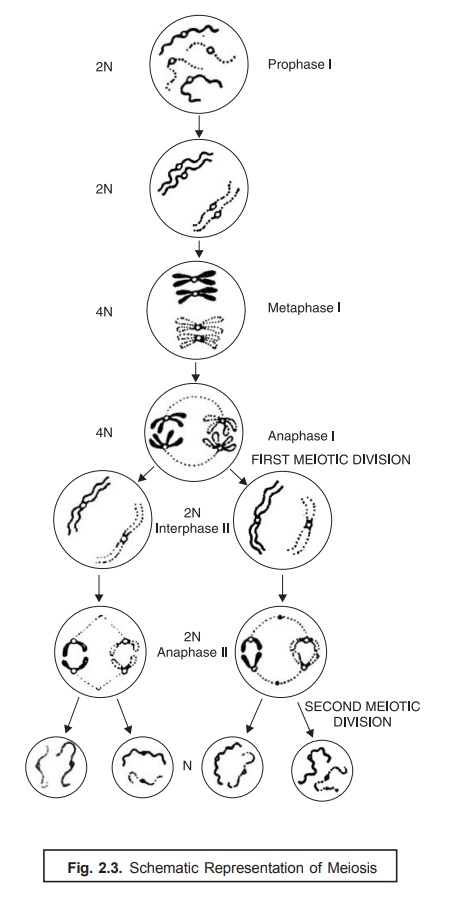
Meiosis. Meiosis refers to a specific process of
two successive cell divisions, giving rise to cells, egg or sperm, that essentially contain half the number of
chromosomes in somatic cells. When fertiliza-tion takes place, the nuclei of
the sperm and ovum fuse and produce a zygote with the full chromosome
complement.
In other
words, the phenomenon of meiosis may
be duly expatiated in sexually reproducing organisms, wherein the prevailing
cellular fusion followed by a reduction in the ‘chromosome number’ is an important and vital feature. The two cells which actually participate in
the sexual reproduction are termed as ‘gametes’,
which fuse to form a ‘zygote’. The
above process is subsequently followed by a nuclear
fusion and the resulting zygote nucleus contains two complete sets of genetic determinants [2N]. In order to adequately maintain the original haploid number in the succeeding
generations, there should be a particular stage at which a definite reduction
in the chromosome number takes place. This process that occurs after the fusion
of gametes is known as meiosis.
Fig. 2.3
illustrates the schematic representation of meiosis, and the various steps
involved may be explained sequentially as follows :
(1) Meiosis
comprises of two meiotic divisions viz., prophase I, and prophase II.
(2) Prophase-I. It represents the first meiotic division, whereby the
homologous chromosomes become
apparently visible as single strands that subsequently undergo pairing.
(3) Each
chromosome renders visible as two
distinct chromatids and thus crossing over takes place.
(4) It is
immediately followed by metaphase I,
wherein the actual orientation of ‘paired
chro-mosomes’ in an equatorial plane and the subsequent formation of a ‘spindle apparatus’ takes place.
(5) It is
followed by Anaphase I, and the homologous centromeres gradually move to the
opposite poles of the spindle.
(6) Telophase-I. It markedly represents the end
of the first meiotic division, and formation of two nuclei takes place.
(8) Interphase-II. Telophase-I
is followed by Interphase-I during which the chromosomes get elongated.
(8) Prophase-II and Metaphase-II. In
prophase-II and metaphase-II the division of centromere and migration of the homologous chromatids occurs, which is duly followed
by anaphase-II, and the desired second meiotic division resulting in the
formation of four haploid* cells.
Eukaryotic Protist. It has
been observed that in several eukaryotic protists belonging to higher ploidy** (> 1) meiosis usually
takes place after the formation of the zygote
and prior to spore formation. In
certain eukaryotes there may even be a critically pronounced alteration of haploid and diploid gen-erations as in the case of the yeast. Interestingly, in this particular instance, the diploid zygote produces a diploid individual that ultimately gives
rise to haploid cells only after
having undergone the phenom-enon of meiosis.
Consequently, the haploid cell may either multiply as a haploid or get fused
with another haploid of the ‘opposite mating
type’ to generate again a diploid.
Example. The life cycle of the eukaryotic
protist may be exemplified by a
typical yeast Saccharomyces cerevisae as
depicted in Fig. 2.4 given below :
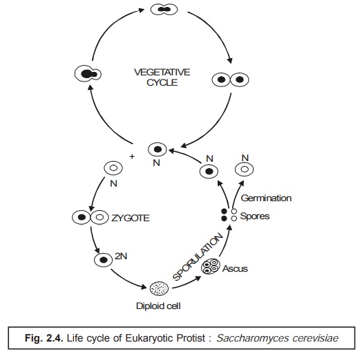
Special Points : There are two cardinal points which, may be borne in mind with regard to the Eukaryotic Protist as stated under :
(i) Despite
of the fact that sexual reproduction could be the only way of reproduction in a
large segment of animals and plants ; it may not be an obligatory event in the
life cycles of many protists.
(ii) In
two glaring situations ; first, protists lacking a sexual stage in
their respective life-cycle ; and secondly,
such species wherein sexuality does exist : the sexual reproduction may be
quite infrequent (i.e., not-so-common).
Important organelles in Eukaryotic Cells : It has
been amply proved and established that the
eukaryotic cells invariably contain certain cytoplasmic organelles other than the nucleus. The important organelles
in eukaryotic cells usually comprise of three
components, namely : mitochondria,
chloroplasts, and the Golgi
apparatus, which shall now be described briefly in the sections that
follows :
Mitochondria. These are mostly found in the
respiring eukaryotes and essentially contain an internal membrane system having characteristic structure and
function. The internal membrane of the mitochondria (cristae) possesses the necessary respiratory electron transport system. The exact number of copies
of mitochondria per cell solely depends upon the cultural parameters and varies
from 1–20 mitochondria per cell. These are generated by the division of the
preexisting organelles containing ribosomes that usually resemble the bacterial
ribosomes. However, the process of protein synthesis in the mitochondria are
very much akin to that in the prokaryotic
cells.
These
cell organelles (rod/oval shape 0.5 μm in
diameter) may be seen by employing a phase-contrast
or electron microscopy. They
mostly contain the enzymes for the
aerobic stages of cell respi-ration and thus are the usual sites of most ATP synthesis chloroplasts [or
Chloroplastids] :
Chloroplasts are found in the photosynthetic
eukaryotic organisms. The
internal membrane of the
chloroplasts is termed as the ‘thylakoid’
which essentially has the three
important components : (a)
photosynthetic pigments, (b) electron
transport system, and (c) photochemical
reaction centres. The number of copies of the chloroplasts depends exclusively
upon the cultural conditions and varies from 40 to 50 chloroplasts per cell.
These are also produced by the division of the preexisting organelles.
Generally,
chloroplasts are the sites of photosynthesis. They possess a stroma and contain
four pigments : chlorophyll a, chlorophyll b, carotene, and xanthophyll.
Golgi Apparatus : The Golgi apparatus is a lamellar membranous organelle invariably
found in the eukaryotic cells and
consists of thickly packed mass of flattened vessels and sacks of different
sizes. The major functions of the Golgi
apparatus are, namely :
·
packaging of both proteinaceous and
nonproteinaceous substances duly synthesized in the endoplasmic reticulum, and
·
their adequate transport to other segments of the
cell.
Golgi apparatus may be best viewed by the aid of electron microscopy. It contains
curved parallel series of flattened
saccules that are often expanded at their ends. In secretory cells, the apparatus
concentrates and packages the secretory product. Its function in other cells,
although apparently impor-tant, is poorly understood.
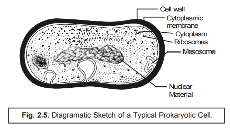
(b) Prokaryotic Cells [‘pro’ = primitive ; ‘karyote’ = nut (refers to nucleus of cell)] :
Prokaryote : is an organism of the kingdom
Monera with a single circular chromosome, without a nuclear membrane, or membrane bound organelles. Included in this
classification are bacteria and cyanobacteria (formerly the blue-green algae)
[SYN : prokaryote].
In fact,
the prokaryotic cell is characterized by the absence of the endoplasmic reticulum (ER) and the cytoplasmic membrane happens to be the
only unit membrane of the cell. If has been observed that the cytoplasmic membrane may be occasionally
unfolded deep into the cytoplasm. An exhaustive electron microscopical studies
would reveal that most prokaryotes {i.e., prokaryotic cells) only two
distinct internal regions, namely :
(a) the cytoplasm ; and (b) the nucleoplasm, as shown in Fig. : 2.5.
Cytoplasm : Cytoplasm refers to
the protoplasm cell outside the nucleus. It is granular in ap-pearance and
contains ribosomes that are specifically smaller in size in comparison to the
corresponding eukaryotic ribosomes.
Nucleoplasm : It refers to the protoplasm of a
cell nucleus. It is fibrillar in character and contains DNA.
With mycoplasmas* as an exception, other prokaryotes invariably comprise of a
defined and rigid cell wall. It has been observed that neither the membranous
structures very much identical to the mitochondria
nor chloroplasts are present in
the prokaryotes. Besides,
the cytoplasmic membrane happens to
be the site of the respiratory electron in the prokaryotes usually. Interestingly, in the photosynthetic micro- organisms (bacteria), the photosynthetic
apparatus is strategically positioned in a particular series of membranous, flattened structures quite
similar in appearance to the thylakoids
; however, these struc-tures are not organized into the respective chloroplasts but are adequately
dispersed in the cytoplasm. Thus, the cytoplasmic membrane contains a plethora
of specific sites for the DNA attachment, and also plays a major role in the
cell division. Here, the cell membrane unlike in the eukaryotic cell does not
generally contain sterols and
polyunsaturated fatty acids (PUFAs). Mostly the fatty acids present are of the saturated type e.g., palmitic acid,
stearic acid etc.
Importantly,
the ‘genetic component’ present in
the prokaryotic cells is
strategically located in the ‘nucleoplasm’ that essentially lacks a defined
nuclear membrane. Nevertheless, it comprises of dou-ble helical DNA without any associated basic proteins. In fact,
the very site of the DNA in prokaryotic protists is much
smaller in comparison to that present in
eukaryotes. In addition, the
prokaryotes do contain
extra-chromosomal DNA, that may replicate autonomously, termed as the ‘plasmids’. How-ever, these can be lost
from the cell without impairment of the ‘cell
viability’. The prokaryotic cells usually exist in a haploid state and
predominently get divided by a process quite identical to mitosis although distinct stages are not recognized so frequently.
A good
number of prokaryotes do possess a cell wall that is vastly different in
composition from that of eukaryotes,
and invariably contains a rather rigid and well-defined polymer termed as the peptidoglycan.* It has been observed
that certain prokaryotes which
essentially possess this aforesaid rigid
structure distinctly exhibit ‘active
movement’ with the help of flagella. Some prokaryotes may also display a ‘gliding
motility’ as could be seen in the ‘blue-green
bacteria’ quite frequently.
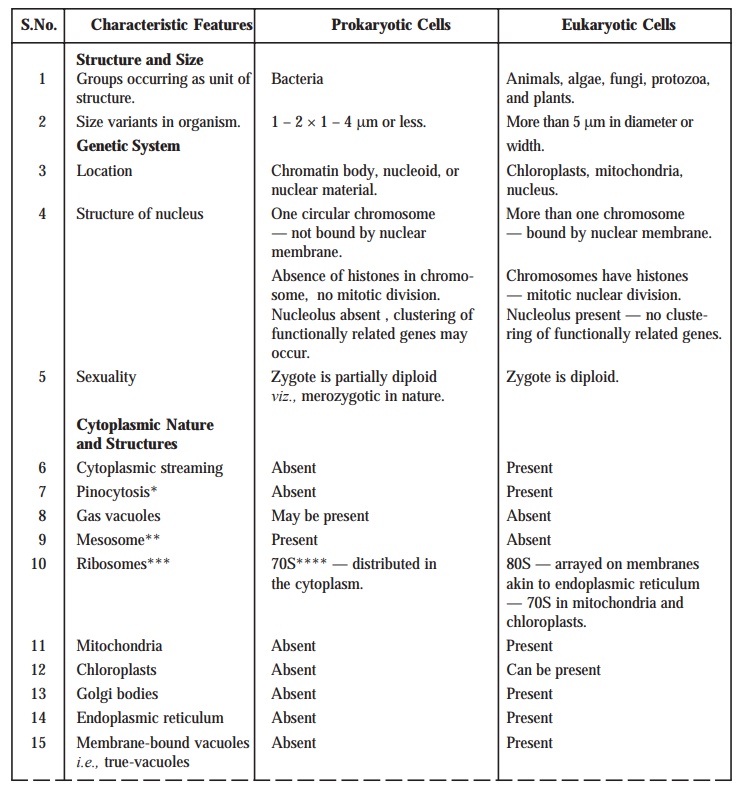
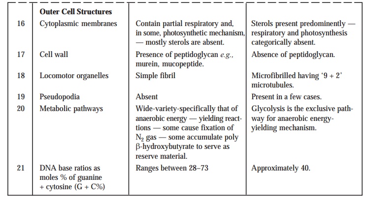
Table : 2.2. records the distinguishing
characteristic features of the
Prokaryotic from the Eukaryotic
Cells.
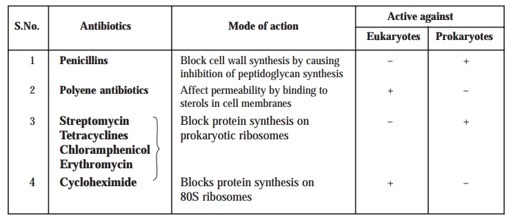
Selective sensitivity to antibiotics. Another
reliable and practical means to differentiate the eukaryotes from prokaryotes
is their characteristic selective sensitivity to certain specific antibiotic(s). However, one may observe that chloramphenicol is toxic only to
bacteria, whereas polyene antibiotics
(e.g., nystatin) bind to sterols in
the cell membranes, and are largely effective exclusively against the eukaryotic protists.
Table 2.3
: summarizes actually the vital and important differences in the activity
against the eukaryotes and prokaryotes with respect to selective
sensitivity to ‘antibiotics’ vis-a-vis their mode of action.
Table 2.3. Differences Between Eukaryotes and Prokaryotes as Regards Selective Sensitivity to Antibiotics/Mode of Action
It is,
however, pertinent to mention here that several cellular functionalities are
prominently and predominently mediated almost differently in these two distinct
types of cells, although the end result is more or less the same.
Related Topics
