Internal Kidney Anatomy
| Home | | Anatomy and Physiology | | Anatomy and Physiology Health Education (APHE) |Chapter: Anatomy and Physiology for Health Professionals: Urinary System
The kidneys are organized into two major regions: an outer renal cortex, which is lighter in color, and an inner renal medulla, which is darker reddish-brown in color.
Internal Kidney Anatomy
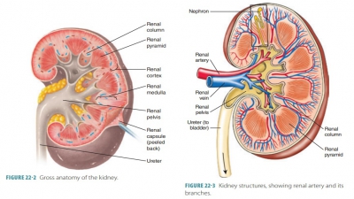
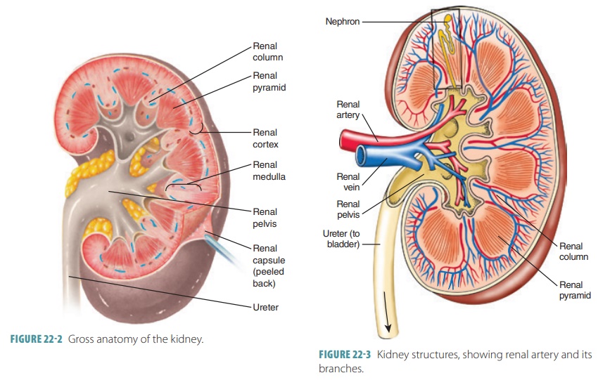
The kidneys are organized into two major regions: an outer renal cortex, which is lighter in color, and an inner renal medulla, which is darker reddish-brown in color (Figure 22-2). Inside the renal sinus, the renal pelvis, a funnel-shaped sac, expands from the supe-rior end of the ureter. It is subdivided into major calyces and minor calyces. Small elevations called renal papillae project into the renal sinus from the renal pelvis walls, and tiny openings leading into the minor calyces pierce each projection. Urine drains continuously from the papillae into the calyces, which empty it to the renal pelvis. It moves through the renal pelvis, into the ureter, and then to the bladder for stor-age. Urine is propelled by peristalsis via the walls of the calyces, pelvis, and uterine smooth muscles.
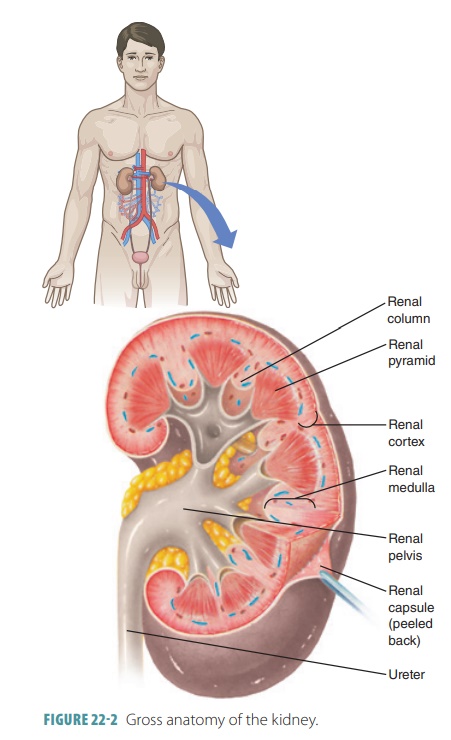
The renal medulla is made of conical tissue called renal pyramids and has striations. Each renal pyr-amid has a broad base that faces the cortex, whereas each apex or papilla faces internally. The renal cortex encloses the medulla, dipping into it between the renal pyramids to form renal columns. The cortex appears to have granules due to tiny tubules associated with the functional units of the kidneys, the nephrons. Each renal pyramid and the cortical tissue that surrounds it make up a lobe of the kidney. Each kidney has approximately eight lobes.
Renal Blood Flow and Nerve Supply
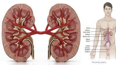
The kidneys are continuously supplied with blood from the renal arteries, which arise from the abdominal aorta. These arteries transport large volumes of blood. While a person rests, the renal arteries carry between 20% and 25% of the total cardiac output, approximately 1,200 mL, into the kidneys every minute.
The renal arteries branch off at right angles inside the kidneys into interlobar arteries, arcuate arteries, and interlobular arteries (FIGURE 22- 3). The final branches of the interlobular arteries lead to the nephrons and are called afferent arterioles. The microscopic blood vessels of the kidney are the main components of how it functions. The right renal artery is the longest, because the aorta lies to the left side of the midline. Nearing a kidney, each renal artery is divided into five segmental arteries. Corresponding, in general, with the arterial pathways, the venous blood returns through a similar series of vessels. Nearly 90% of the blood that enters the kidney perfuses the renal cortex. The arcuate artery lies on the boundary between the cortex and medulla of the kidneys.
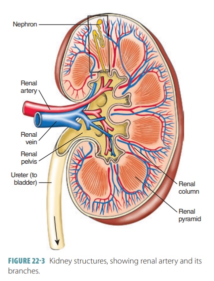
The renal vein then joins the inferior vena cava. Venous blood returns through a series of vessels that correspond generally to arterial pathways. Blood that leaves each kidney flows from the cortex through the cortical radiate vein, arcuate vein, interlobar vein, and finally, the renal vein. Unlike renal arteries, there is no segmental component in the vein system. Exiting the kidney, the renal veins empty to the inferior vena cava. The renal plexus provides the kidney’s nerve supply and also the nerve supply of the ureter. The renal plexus is a varied network of autonomic ganglia and nerve fibers that derives from the celiac plexus. It has many sympathetic fibers as its supply, from the inferior thoracic and first lumbar splanchnic nerves. These nerves trace the renal artery and are sympa-thetic vasomotor fibers regulating renal blood flow via adjustment of renal arteriole diameter. They also influence the nephrons’ urine formation.
1. Describe the anatomy, location, and structure of the kidneys.
2. Differentiate between the renal cortex, medulla, and pelvis.
3. From where do the renal arteries arise and into what structure does a renal vein drain?
4. Explain the blood flow through a kidney.