Hairs
| Home | | Anatomy and Physiology | | Anatomy and Physiology Health Education (APHE) |Chapter: Anatomy and Physiology for Health Professionals: Support and Movement: Integumentary System
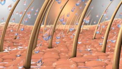
Hairs (pili) project above the skin surface over most of the body, except for the sides and soles of the feet, the palms of the hands, the sides of the fingers and toes, the lips, and parts of the external genitalia.
Hairs
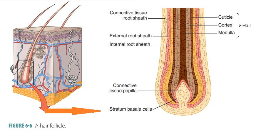
Hairs (pili) project above the skin surface over most of the body, except for the sides and soles of the feet, the palms of the hands, the sides of the fingers and toes, the lips, and parts of the external genitalia. They begin to form during embryologic development and are also known as epidermal derivatives because they arise from the epidermis. There are about 2.5 million hairs on the human body, of which over 75% is on the general body surface and not the head. Hairs are structures produced in organs called hair follicles (FIGURE 6-6). They consist of a large amount of dead keratinized cells, dominated by hard keratin. Hair follicles extend from the skin surface into the dermis, containing hair roots that are nourished with dermal blood. Each hair follicle is attached to an arrector pili muscle, which helps the hair shaft (in which keratinization is complete) to stand on end when it contracts. This occurs during emotional upset and cold temperatures, causing gooseflesh or goose bumps. Hairs are pushed upward as epidermal hair cells divide and grow, becoming keratinized and then dying.
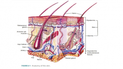
Each hair follicle is folded from
the epidermal sur-face into the dermis. They may extend into the hypo-dermis of
the scalp. Each follicle originates at about 4 mm below the skin surface,
expanding to form a hair bulb. A root hair plexus or hair follicle receptor
con-sists of a cluster of sensory nerve
endings, wrapping around each hair bulb. When the hair is bent, these endings
are stimulated, meaning that hairs act as touch receptors with extreme
sensitivity. Nipple-like dermal tissue
makes up a hair papilla, which
protrudes into each hair bulb. It contains a knot of capillaries that give
nutrients to the growing hair. A fibrous
peripheral connective tissue sheath makes
up the wall of a hair fol-licle. This derives from the dermis. The other
compo-nents of the hair follicle wall are the thickened basal glassy membrane and the inner epithelial root sheath. This sheath becomes thinner as it
approaches the hair bulb, with only one layer of epithelial cells covering the
papilla.
The hair
matrix is the actively dividing part of
the hair bulb that produces the hair. It originates in the hair bulge, just a small portion of 1 mm above the hair bulb.
Chemical signals that reach the hair bulge cause certain cells to move to the
papilla, divide, and produce new hair cells. The older part of each hair is
then pushed upward, with the fused cells getting more keratin and dying.
Each hair has three concentric
layers known as the medulla, cortex,
and cuticle. The central core of a
hair is the medulla. It is made up of air spaces and large cells. The medulla
is the only hair portion that contains soft keratin. It does not exist in fine
hairs.

Surrounding the medulla is the
bulky cortex, made up of a few layers
of flat cells. A single layer of cells forms the outermost cuticle of a hair. In the cuticle, cells are overlapped like
shingles, which helps keep each hair shaft from matting with others. Most hard
keratin is in the cuticle, which provides strength to the hair shaft and keeps
the inner layers compacted tightly together. The cuticle experiences the most
abrasion and usually wears away at the tip. This causes what is commonly known
as split ends, in which fibrils of
keratin from the cortex and medulla become split. The rough surfaces of a hair
cuticle are smoothed by hair conditioning products, which give a shiny
appearance to a person’s head of hair.
Hair color is reflected by
genetics and variations in the pigment produced by melanocytes at the hair
papilla. Darker hair has more eumelanin (which is brownish-black), whereas
lighter hair has more phe-omelanin (which is reddish-yellow). The different
forms of melanin give hair a wide variety of shades, ranging from dark brown to
yellow-brown to red. Albinos have white hair because their hair shafts
com-pletely lack melanin. Hormonal and environmental factors also influence the
hair’s condition. As pigment production decreases with age, hair color
lightens. White hair results from a lack of pigment along with the presence of
air bubbles in the medulla of the hair shaft. As the proportion of white hairs
increases, the overall hair color is described as gray.
Hairs are basically classified as
either vellus or terminal. Adult women and newborn children have body hair known as vellus
hair, which is fine and pale in color. Terminal
hair is coarser and longer, found on the scalp and eyebrows, and often
darker than vellus hair. Terminal hairs appear in the axillary and pubic
regions of males and females during puberty. On males at puberty, they also
appear on the face, chest, and usually the arms and legs. These hairs are
stimulated to grow by androgens (primarily tes-tosterone). Large amounts of
male hormones cause thick terminal hair growth. Nutrition and hormones
influence hair density and growth. Chronic physical inflammation or irritation
may cause increased local hair growth. Hair growth that is not cosmetically
attractive may be slowed or stopped by electrolysis or laser treatments.
Hair grows at an average rate of
2.5 mm per week, but this varies with sex, age, and body regions. Growth cycles
occur in each hair follicle, including an active phase that ranges from weeks
to years and a regres-sive phase when hair matrix cells die. The hair folli-cle
base and hair bulb then shrink, causing the hair papilla to be moved upward to
touch the part of the follicle that does not regress. Then, a 1–3-month
rest-ing phase occurs. After this, the cycling area of the follicle is
regenerated. Activated bulge cells migrate to the papilla. The matrix is then
able to form a new hair. Many proteins control a hair’s life span, and scalp
hair follicles are active for up to 10 years before being inac-tivated for
several months. Approximately, 90 scalp hairs are lost per day. The eyebrows
never reach the length of the scalp hair because each eyebrow follicle is
active for only 3–4 months.
Each hair follicle of the body
has a limited num-ber of growth cycles, with growing being fastest between the
teenage years and the 40s. Hair thinning occurs after this time, because
shedding happens more quickly than hair replacement. Both sexes, beginning in
middle age, experience a certain degree of hair thinning and/or alopecia (baldness), but it is more
commonly seen in men. By age 35, approx-imately 40% of men have visible hair
loss, and by age 60 the percentage is about 85%. Scalp hair loss usually begins
at the anterior hairline, progressing posteriorly. The hair becomes thinner as vellus
hairs being to replace the coarser terminal hairs. However, true baldness (most commonly, male pattern bald-ness ) is not the
same situation. It is sex influenced and
genetically determined, linked to a gene that activates during adulthood and
changes how the hair follicles respond to dihydrotestosterone, which is a
metabolite of testosterone.
1. Describe
the mechanism that causes hairs to “stand on end” and also causes goose bumps.
2. Explain
the tissue that comprises the hair papilla.
3. Describe
the hair follicles, the root hair plexus, and the hair matrix.