Gprotein Coupled Receptors (GPCR)
| Home | | Pharmacology |Chapter: Essential pharmacology : Pharmacodynamics Mechanism Of Drug Action; Receptor Pharmacology
These are a large family of cell membrane receptors which are linked to the effector (enzyme/ channel/carrier protein) through one or more GTPactivated proteins (Gproteins) for response effectuation.
G-PROTEIN COUPLED RECEPTORS (GPCR)
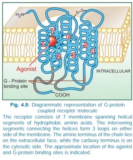
These are a large family of cell membrane receptors which are linked to the effector (enzyme/ channel/carrier protein) through one or more GTPactivated proteins (Gproteins) for response effectuation. All such receptors have a common pattern of structural organization (Fig. 4.5). The molecule has 7 αhelical membrane spanning hydrophobic amino acid (AA) segments which run into 3 extracellular and 3 intracellular loops. The agonist binding site is located somewhere between the helices on the extracellular face, while another recognition site formed by cytosolic segments binds the coupling Gprotein. The Gproteins float in the membrane with their exposed domain lying in the cytosol, and are heterotrimeric in composition (α, β and γ subunits). In the inactive state GDP is bound to their exposed domain; activation through the receptor leads to displacement of GDP by GTP. The active αsubunit carrying GTP dissociates from the other two subunits and either activates or inhibits the effector. The βγ subunits have also been shown to modulate certain effectors like receptoroperated K+ channels, adenylylcyclase (AC) and phospholipase C.
A number of G proteins distinguished by their α subunits have been described. The important ones with their action on the effector are:
Gs : Adenylyl cyclase ↑, Ca2+ channel ↑
Gi : Adenylyl cyclase ↓, K+ channel ↑
Go : Ca2+ channel ↓
Gq : Phospholipase C ↑
G13 : Na+/H+ exchange ↑
In addition Gn, Gk, Gt and Golf have been distinguished. A limited number of Gproteins are shared between different receptors and one receptor can utilize more than one Gprotein (agonist pleotropy), e.g. the following couplers have been associated with different receptors.
Receptor Coupler
Muscarinic Gi, Go, Gq
Dopamine D2 Gi, Go
βadrenergic Gs, Gi
α1adrenergic Gq
α2adrenergic Gi, Gs, Go
GABAB Gi, Go
5HT Gi, Gq, Gs, Gk
In addition, a receptor can utilize different biochemical pathways in different tissues.
The α-subunit has GTPase activity: the bound GTP is slowly hydrolysed to GDP: the αsubunit then dissociates from the effector to rejoin its other subunits, but not before the effector has been activated/inhibited for a few seconds and the signal has been amplified. The onset time of response through this type of receptors is also in seconds.
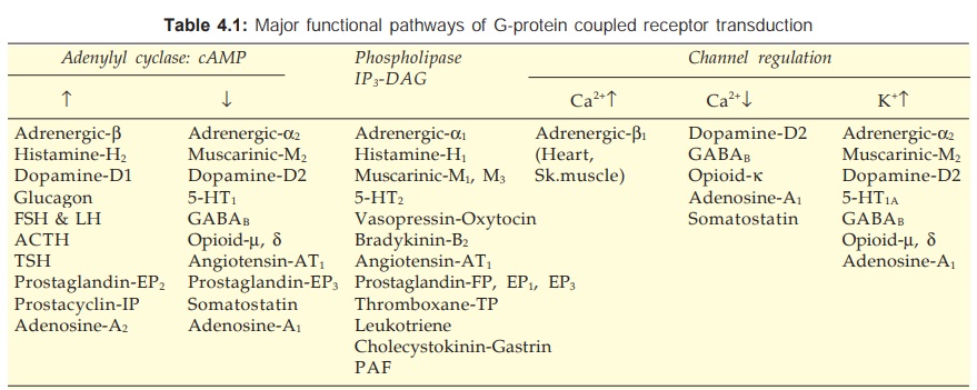
There are three major effector pathways (Table 4.1) through which GPCRs function.
a) Adenylyl cyclase: cAMP pathway Activation of AC results in intracellular accumulation of second messenger cAMP (Fig. 4.6) which functions mainly through cAMPdependent protein kinase (PKA). The PKA phosphorylates and alters the function of many enzymes, ion channels, transporters and structural proteins to manifest as increased contractility/impulse generation (heart), relaxation (smooth muscle), glycogenolysis, lipolysis, inhibition of secretion/mediator release, modulation of junctional transmission, hormone synthesis, etc. In addition, cAMP directly opens a specific type of membrane Ca2+ channel called cyclic nucleotide gated channel (CNG) in the heart, brain and kidney. Responses opposite to the above are produced when AC is inhibited through inhibitory G-protein.

b) Phospholipase C: IP3DAG pathway Activation of phospholipase C (PLc) hydrolyses the membrane phospholipid phosphatidyl inositol 4, 5bisphosphate (PIP2) to generate the second messengers inositol 1,4,5trisphosphate (IP3) and diacylglycerol (DAG). The IP3 mobilises Ca2+ from intracellular organellar depots and DAG enhances protein kinase C (PKc) activation by Ca2+ (Fig. 4.7). Cytosolic Ca2+ (third messenger in this setting) is a highly versatile regulator acting through calmodulin (CAM), PKc and other effectors—mediates/modulates contraction, secretion/transmitter release, eicosanoid synthesis, neuronal excitability, intracellular movements, membrane function, metabolism, cell proliferation, etc. Like AC, the PLc can also be inhibited through inhibitory Gprotein when directionally opposite responses would be expected.
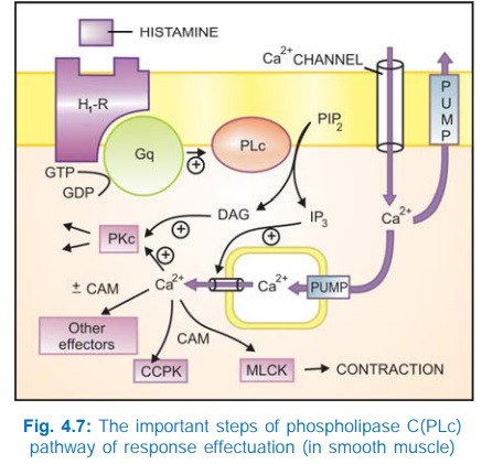
Intracellular Ca2+ release has been found to occur in waves (Ca2+ mediated Ca2+ release from successive pools facilitated by inositol 1, 3, 4, 5tetrakisphosphate—IP4) and exhibits a variety of agonist and concentration dependent oscillatory patterns. The activation of different effectors may depend on the amplitude and pattern of these oscillations. Thus, the same intracellular messenger can trigger different responses depending on the nature and strength of the extracellular signal.
c) Channel regulation The activated Gproteins can also open or close ionic channels specific for Ca2+, K+ or Na+, without the intervention of any second messenger like cAMP or IP3, and bring about hyperpolarization/depolarization/ changes in intracellular Ca2+. The Gs opens Ca2+ channels in myocardium and skeletal muscles, while Gi and Go open K+ channels in heart and smooth muscle as well as close neuronal Ca2+ channels. Physiological responses like changes in inotropy, chronotropy, transmitter release, neuronal activity and smooth muscle relaxation follow. Receptors found to regulate ionic channels through Gproteins are listed in Table 4.1.
Related Topics
