Dermis - Skin
| Home | | Anatomy and Physiology | | Anatomy and Physiology Health Education (APHE) |Chapter: Anatomy and Physiology for Health Professionals: Support and Movement: Integumentary System
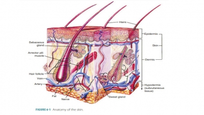
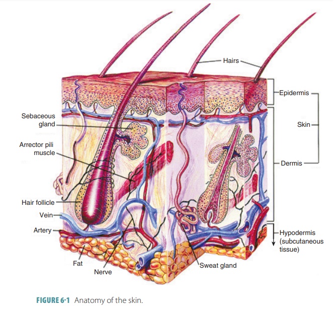
The dermis lies between the epidermis and the sub-cutaneous layer and has two major components: a superficial papillary layer and a deeper reticular layer.
Dermis
The dermis lies between the epidermis and the sub-cutaneous layer and
has two major components: a superficial papillary layer and a deeper reticular
layer. The dermis also contains all cells of connective tissue proper.
Epidermal accessory organs extend into the dermis, and both the papillary and
reticular layers of the dermis contain many blood vessels, lymph vessels, and
nerve fibers. The dermis is the second major skin structure and is a strong and
flexible connective tis-sue. Dermal cells contain fibroblasts, macrophages, and
smaller amounts of mast cells and white blood cells. The major portions of hair
follicles, oil glands, and sweat glands are found in the dermis, even though
they derive from the epidermal tissue.
The papillary
layer consists of areolar tissue and
contains capillaries, lymphatics, and sensory neurons. The papillary layer is
named for the dermal papillae that project between the epidermal ridges. It is
a thin and superficial areolar connective tissue with interwo-ven collagen and
elastic fibers. The papillary layer is loose, allowing phagocytes and various
defensive cells to move freely, searching for bacteria that have gotten through
the skin. Dermal papillae often contain loops of capillaries or may contain
pain receptors and touch receptors. In the thicker skin of the palms or soles,
these papillae are above larger dermal
ridges, which then cause the epidermis to form its epidermal ridges. These ridges leave pressure marks that are
commonly referred to as fingerprints,
unique to every individual human being.
The deeper reticular layer is made up of connec-tive tissue containing
collagen and elastic fibers. The boundary between the papillary and reticular
layers is not distinct. The reticular layer makes up approxi-mately 80% of the
overall thickness of the dermis. It is nourished by the cutaneous plexus. Collagen fibers mostly run parallel to the skin
surface, with less dense regions known as separations
forming cleavage lines in the skin. Also known as tension
lines, these lines are used for surgeries to make parallel incisions,
meaning better healing afterward. A third type of skin marking, flexure lines, occur close to joints, where the dermis is more tightly
secured to deeper structures. Examples include the creases on the palms of the
hands. In the papillary layer, small arteries form a branched net-work known as
the papillary plexus, which provides
arterial blood to the capillaries along the epidermis– dermis boundary.
The collagenous and elastic
fibers of the dermis make it both tough and elastic. The skin’s water content
helps it to be flexible and resilient, which is known as skin turgor. A sign of dehydration is the loss of skin turgor. Processes from nerve cells are
located through-out the dermis. Motor processes carry impulses to the dermal
glands and muscles, whereas sensory processes carry impulses back to the brain
and spinal cord. Cuta-neous sensations include touch, hot, cold, and pain.
1. Describe
the layer of the dermis that determines a person’s fingerprints.
2. What
forms cleavage lines in the skin?
3. What
are the functions of the dermis?