Classification of Nerve Fibers
| Home | | Anatomy and Physiology | | Anatomy and Physiology Health Education (APHE) |Chapter: Anatomy and Physiology for Health Professionals: Control and Coordination: Neural Tissue
Nerve fibers are classified by their diameter, degree of myelination, and speed of conduction. There are three primary groups of nerve fibers:
Classification
of Nerve Fibers
Nerve fibers are classified by
their diameter, degree of myelination, and speed of conduction. There are three
primary groups of nerve fibers:
■■ Group A fibers: These mostly
serve the joints, skeletal muscles, and skin, and are primarily somatic sensory
and motor fibers, with the largest diameter of all types of fibers and thick
myelin sheaths. These fibers conduct impulses at speeds as high as 300 miles
per hour.
■■ Group B fibers: Of
intermediate diameter, with light myelination, group B fibers conduct impulses
at speeds averaging approximately 30 miles per hour.
■■ Group C fibers: These fibers
are nonmyelinated with the smallest diameter and cannot create saltatory
conduction; they conduct impulses at 2 miles per hour or less.
Both B and C fibers include motor
fibers of the ANS that serve the smaller somatic sensory fibers that transmit
sensory impulses from the skin (including small touch and pain fibers),
visceral sensory fibers, and those that serve the visceral organs.
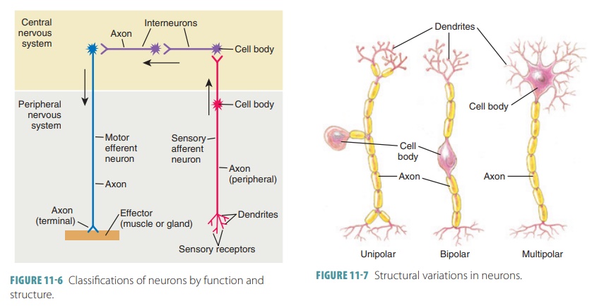
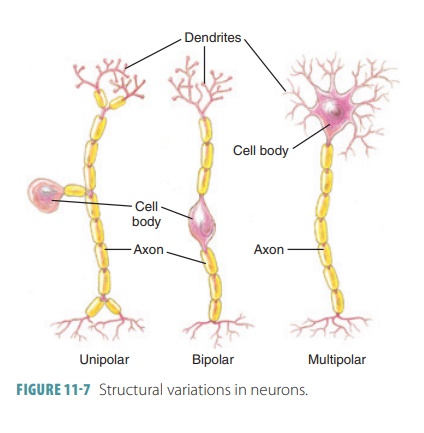
Structural Classification of Neurons
Neurons are classified based on
the number of pro-cesses that extend from their cell bodies. The three major
structural categories of neurons are multipolar, bipolar, and unipolar:
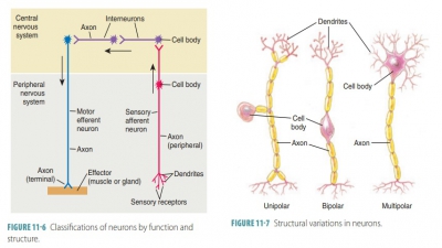
■■ Multipolar neurons: They make
up most of the neurons whose cell
bodies lie within the brain or spinal cord. They have three or more processes
that arise from their cell bodies, with only one process being an axon and the
rest being den-drites. Multipolar neurons are the most common, and more than
99% of neurons in the human body are multipolar (FIGURE
11-6). They are also the most common
type in the CNS.
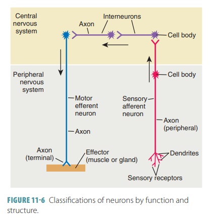
■■ Bipolar neurons: These
neurons exist only in specialized parts of the eyes and nose. They have only
two processes arising from their cell bodies. Only one process of each neuron
is an axon and the other is a dendrite. Bipolar neurons are very rare in the
body. They are located in the retinas of the eyes and in the nasal cavity.
■■ Unipolar neurons: Often
aggregated in specialized ganglia located outside the brain and spinal cord,
these neurons have a single short process extending from the cell body that
divides into two T-like branches that function more like a single axon. One
branch of the more distal peripheral process is associated with dendrites near a
peripheral body part and the other branch (the central
process) enters the brain or spinal
cord. Unipolar neurons originate as bipolar neurons and are more accurately described
as pseudounipolar neurons.
Functional Classification of Neurons
The functional classification of
neurons is based on the direction in which action potentials are conducted:
■■ Sensory neurons (afferent
neurons): They carry nerve
impulses from the peripheral body parts into the CNS. They may have receptor
ends at the tips of the dendrites or receptor cells that are associated with
the dendrites in the sensory organs or the skin (FIGURE
11-7). Somatic sen-sory neurons monitor the external environment, whereas visceral sensory neurons monitor the body’s internal environment.
Sensory receptors are classified as interoceptors, exteroceptors, and
proprioceptors. Interoceptors provide sen-sations of deep pressure, distension, and pain and are
found in the digestive, cardiovascular, respiratory, reproductive, and urinary
systems. Exteroceptors provide perception of tempera-ture, touch, pressure, smell,
taste, equilibrium, hearing, and sight. Proprioceptors provide per-ception of skeletal muscle and joint movement
and position.
■■ Interneurons: They conduct action potentials from one neuron to another within the CNS. The cell bodies
of some interneurons form masses called nuclei in the CNS, which are similar
toganglia.
■■ Motor neurons (efferent neurons): They conduct action potentials away from the CNS toward mus-cles or glands. Somatic motor neurons innervate skeletal muscles. Their cell bodies lie within the CNS, whereas their axons extend outward within peripheral nerves, innervating skeletal muscle fibers at the neuromuscular junctions. Visceral motor neurons innervate smooth and cardiac muscle, glands, and adipose tissue. The axons of these neurons that lie within the CNS innervate other visceral motor neurons in the peripheral autonomic ganglia. Visceral motor neurons that have cell bodies in these ganglia innervate and control the peripheral effectors. Axons extend-ing from the CNS to an autonomic ganglion are known as preganglionic fibers. Axons that connect ganglion cells to peripheral effectors are known as postganglionic fibers.
1. Describe
the structures of neurons, dendrites, and axons.
2. Identify
the differences between sensory and motor neurons.
3. What
are the three major structural categories ofneurons?
4. Differentiate
between multipolar and bipolar neurons.