Cholinoceptors
| Home | | Pharmacology |Chapter: Essential pharmacology : Cholinergic System And Drugs
Two classes of receptors for ACh are recognised —muscarinic and nicotinic; the former is a G protein coupled receptor, while the latter is a ligand gated cation channel.
CHOLINOCEPTORS
Two
classes of receptors for ACh are recognised —muscarinic and nicotinic; the
former is a G protein coupled receptor, while the latter is a ligand gated
cation channel.
Muscarinic
These receptors are
selectively stimulated by
muscarine and blocked by atropine. They are located primarily on autonomic
effector cells in heart, blood vessels, eye, smooth muscles and glands of
gastrointestinal, respiratory and urinary tracts, sweat glands, etc. and in the
CNS. Subsidiary muscarinic receptors are also present in autonomic ganglia
where they appear to play a modulatory role by inducing a longlasting late
EPSP.
Muscarinic autoreceptors are present prejunctionally on postganglionic cholinergic nerve endings: their activation inhibits further ACh release. Similar ones have been demonstrated on adrenergic terminals: their activation inhibits NA release (may contribute to vasodilator action of injected ACh). All blood vessels have muscarinic receptors (though most of them lack cholinergic innervation) located on endothelial cells whose activation releases EDRF which diffuses to the smooth muscle to cause relaxation.
Subtypes of Muscarinic Receptor
By pharmacological as
well as molecular cloning techniques, muscarinic receptors have been divided
into 5 subtypes M1, M2, M3, M4 and
M5. The first 3 are the major subtypes (Table 7.3) that are present
on effector cells as well as on prejunctional nerve endings, and are expressed
both in peripheral organs as well as in the CNS. The M4 and M5
receptors are present mainly on nerve endings in certain areas of the brain and
regulate the release of other neurotransmitters. Functionally, M1, M3
and M5 fall in one class while M2 and M 4 fall
in another class. Muscarinic agonists have shown little subtype selectivity,
but antagonists (pirenzepine for M1, tripitramine for M2
and darifenacin for M3) are more selective. Most organs have more
than one subtype, but usually one subtype predominates in a given tissue.
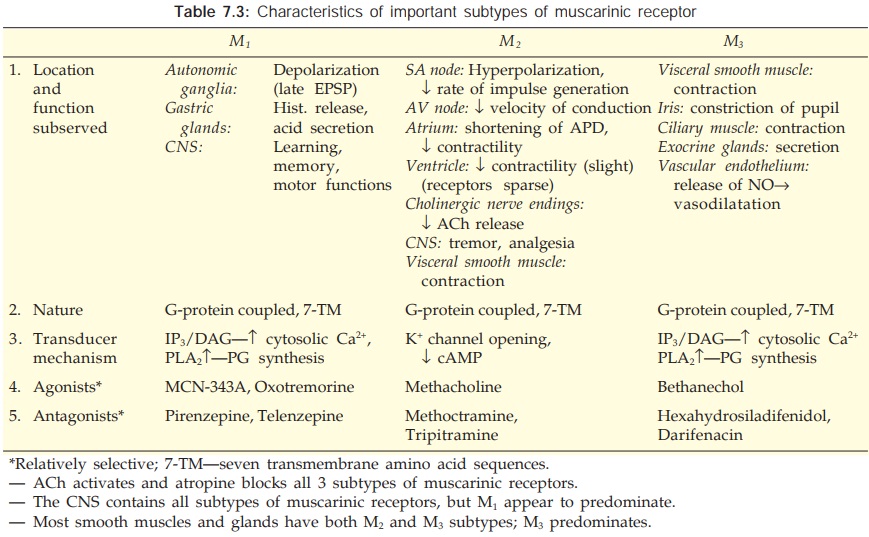
M1: The M1 is primarily a neuronal receptor located on ganglion cells and central
neurones, especially in cortex, hippocampus and corpus striatum. It plays a
major role in mediating gastric secretion, relaxation of lower esophageal
sphincter (LES) on vagal stimulation and in learning, memory, motor functions,
etc.
M2: Cardiac muscarinic
receptors are predominantly M2 and mediate vagal bradycardia.
Autoreceptors on cholinergic nerve endings are also of M2 subtype.
Smooth muscles express some M2 receptors as well which, like M3,
mediate contraction.
M3: Visceral smooth muscle
contraction and glandular secretions
are elicited through M3 receptors, which also mediate vasodilatation
through EDRF release. Together the M2 and M3 receptors
mediate most of the wellrecognized muscarinic actions including contraction of
LES.
The muscarinic receptors
are G-protein coupled receptors having the characteristic 7 membrane traversing
amino acid sequences. The M1 and M3 (also M5)
subtypes function through Gq protein and activate membrane bound phospholipase
C (PLc)—generating inositol trisphosphate (IP3) and diacylglycerol
(DAG) which in turn release Ca2+ intracellularly—cause
depolarization, glandular secretion and raise smooth muscle tone. They also
activate phospholipase A2 resulting in enhanced synthesis and
release of prostaglandins and leucotrienes in certain tissues. The M2
(and M4) receptor opens K+ channels (through βγ subunits of
regulatory protein Gi) and inhibits adenylyl cyclase (through α subunit of Gi)
resulting in hyperpolarization, reduced pacemaker activity, slowing of conduction
and decreased force of contraction in the heart. The M4 receptor has
been implicated in facilitation/ inhibition of transmitter release in certain
areas of the brain, while M5 has been found to facilitate dopamine
release and mediate reward behaviour.
Nicotinic
These receptors are
selectively activated by nicotine and blocked by tubocurarine or hexamethonium.
They are rosettelike pentameric structures (see
Fig. 4.4) which enclose a ligand gated cation channel: their activation causes
opening of the channel and rapid flow of cations resulting in depolarization
and an action potential. On the basis of location and selective agonists and
antagonists two subtypes NM and NN (previously labelled N1
and N2) are recognized (Table 7.4).
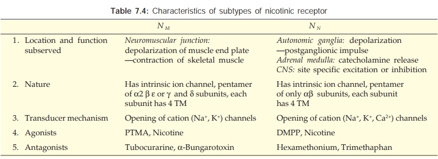
NM: These are present at
skeletal muscle endplate: are selectively
stimulated by phenyl trimethyl ammonium (PTMA) and blocked by tubocurarine.
They mediate skeletal muscle contraction.
NN: These are present on
ganglionic cells (sympathetic as well as parasympathetic), adrenal medullary
cells (embryologically derived from the same site as ganglionic cells) and in
spinal cord and certain areas of brain. They are selectively stimulated by
dimethyl phenyl piperazinium (DMPP), blocked by hexamethonium, and constitute
the primary pathway of transmission in ganglia.
Related Topics
